Friday, 26 February 2016

Polynucleotides & DNA

Nucleotides --> Polynucleotides

  • The Nucleotides Join Together between the Phosphate group of one nucleotide, and between a Sugar of another. This forms a Phosphodiester Bond (made up of the phosphate group and two ester bonds - Phospho-Di-Ester)
  • The chain of Sugars and Phosphates is known as the Sugar-Phosphate Backbone
  • Polynucleotides can be Broken Down into Nucleotides again by Breaking the Phosphodiester Bonds.
Double Helix

  • Two Polynucleotides join together by Hydrogen Bonding Between the Bases.
  • Each base can only bond with a particular partner: this is called Complimentary Base Pairing.
  • Adenine (A) always pairs with Thymine (T) and Cytosine (C) always pairs with Guanine (G) (I remember it as the straight letters go together, and the curly letters go together)
  • A Pyrimidine always pairs with a Purine.
  • 2 Hydrogen bonds form between A-T and 3 Hydrogen Bonds form between G-C
  • Two Antiparallel (going in opposite directions) polynucleotide Strands Twist to form the DNA Double Helix
Purifying DNA - Precipitation Reaction
  1. Break Up Cells using a blender
  2. Make a mixture of washing up liquid, salt, and distilled water, this mixture is called the Detergent
  3. Add broken cells to a beaker containing the detergent
  4. Put the beaker in a Water Bath (60°C) for 15 Minutes.
  5. Now, put the beaker in an Ice Bath, then Filter the mixture.
  6. Transfer a small amount of the filtered mixture to a clean boiling tube.
  7. Add Protease Enzymes
  8. Slowly Dribble some cold ethanol down the side of the boiling tube so it forms a Layer on top of the mixture.
  9. Leave it for a few minutes and a White Precipitate should form, which can be removed with a Glass Rod.
Self-Replication
DNA can Copy Itself before cell division
  1. DNA  Helicase (Enzyme) Breaks the Hydrogen Bonds Between Two polynucleotide DNA Strands - The helix Unzips.
  2. Each Original Strand becomes a Template for a new strand. Free-Floating DNA Nucleotides Join to the exposed bases of each original strand by Complementary Base Pairing.
  3. The Nucleotides of the New Strand are joined together by DNA Polymerase (Another Enzyme). This forms the Sugar-Phosphate Backbone. Hydrogen Bonds Form between the bases on the Original and New Strand. It then Twists to form a Double Helix.
  4. Each new DNA molecule contains One strand from the Original DNA Molecule and One New Strand

Nucleotides


  • A Nucleotide is yet another type of Biological Molecule, it's made from a Pentose Sugar, a Nitrogenous Base and a Phosphate Group.
  • All Nucleotides contain the elements, Phosphorus, Oxygen, Nitrogen, Carbon and Hydrogen 


  • Nucleotides are the Monomers that make up DNA and RNA
  • ADP and ATP are Special types of Nucleotides used to Store and Transport Energy in cells

Deoxyribose (Shown Above)

  • The pentose sugar in a DNA nucleotide is 'Deoxyribose'
  • Each DNA Nucleotide has the Same Sugar and Phosphate Group, it is the Base on each DNA nucleotide that Varies.
  • There are 4 possible bases, Adenine (A), Thymine (T), Guanine (G) and Cytosine (C)
  • Adenine and Guanine are a type of base called Purine.
  • Thymine and Cytosine are a type of base called Pyrimidine.


  • Purine: contains 2 Carbon-Nitrogen Rings joined together
  • Pyrimidine: Only 1 Carbon-Nitrogen Ring - so it is Smaller than purine.
  • A Molecule of DNA contains Two Polynucleotide Chains - each chain having lots of nucleotides joined together.
Ribose 
  • RNA contains Nucleotides with a Ribose sugar instead of deoxyribose.
  • An RNA molecule also has a Phosphate group, and one of four different bases.
  • However, in RNA, Uracil (U), which is a Pyrimidine, Replaces Thymine as a base.
  • An RNA molecule is made up of a Single Polynucleotide Chain

ADP and ATP
  • To Phosphorylate a nucleotide, you Add One or More Phosphate Groups to it.
  • ADP (Adenosine Diphosphate) contains the base Adenine, the sugar Ribose and Two Phosphate Groups.
  • ATP (Adenosine Triphosphate) contains the base Adenine, the sugar Ribose, and Three Phosphate Groups.
ADP, ATP and Energy
  • ATP provides Energy for Chemical Reactions in the Cell.
  • ATP is Synthesised from ADP and Inorganic Phosphate (Pi) using the Energy From an Energy-Releasing Reaction, (e.g. the breakdown of glucose in respiration.
  • The ADP is Phosphorylated to form ATP and a Phosphate Bond is formed.
  • Energy is Stored in the Phosphate Bond. When this Energy is Needed by a cell, ATP is Broken Back Down into ADP and Inorganic Phosphate (Pi). The Energy is Released from the Phosphate Bond and Used by the Cell.

Thursday, 25 February 2016

Biochemical Tests and Separating Molecules

Biosensors
A Biosensor is a device that uses a Biological Molecule, such as an Enzyme to detect a Chemical.
The biological molecule produces a Signal (i.e. a chemical  signal) which is Converted into an Electrical Signal by a Transducer.
The electrical signal is then processed and can be used to work out other information.

Example:
A Glucose Biosensor is used to determine the Concentration of Glucose in a Solution.
It does this using the enzyme 'Glucose Oxidase' and Electrodes.
The enzyme Catalyses the Oxidation of glucose at the Electrodes. this creates a Charge, which is Converted into an Electrical Signal by the Electrodes (Transducer)
The Electrical Signal is then Processed to work out the Initial Glucose Concentration

Chromatography
The main use is Separating.
Once a solution is separated, we can Identify the Components.
It can be used to Identify Biological Molecules such as Amino Acids, Carbohydrates, Vitamins and Nucleic Acids.
There are loads of different types of chromatography, but we only need to know two, Paper Chromatography and Thin-Layer Chromatography.

Both methods of chromatography have two basic phases:
A MOBILE PHASE

  • Where the Molecules Can Move
  • in both paper and thin-layer chromatography, the Mobile Phase is a Liquid Solvent, such as Ethanol or Water.
A STATIONARY PHASE
  • Where the Molecules Can't Move
  • In Paper chromatography, the stationary phase is a piece of Paper.
  • In Thin-Layer Chromatography, the stationary phase is a Thin (<0.5mm) Layer of a Solid, i.e. Silica Gel, Glass or Plastic.
They both use the same basic method:
  1. The Mobile Phase moves Through or Over the Stationary Phase, 
  2. The Components in the mixture spend Different amounts of Time in the Mobile phase And the Stationary phase.
  3. The Components that spend Longer in the Mobile phase travel Faster or Further.
  4. The time spent in the different phases is what separates out the components of the mixture.
PAPER CHROMATOGRAPHY
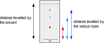
  1. Draw a pencil line near the bottom of the chromatography paper and put a concentrated spot of the mixture of amino acids on it.
  2. Add a small amount of prepared Solvent in a beaker and Dip the bottom of the paper into it. Cover it with a lid to stop the solvent evaporating.
  3. As the solvent spreads up the paper, the different amino acids move with it, but at different rates, so they separate out.
  4. When the solvent's nearly reached the top, Take the paper Out and Mark the Solvent Front (the highest point the solvent has reached), now leave the paper to Dry before analysing it.
  5. Since amino acids are colourless, Spray them with Ninhydrin solution to turn the amino acids purple, then use the Rf values to Identify the separated molecules.


Colorimetry

Colorimetry is used to determine the Concentration of a Glucose Solution.

  • To do this, we need to use Benedict's Reagent and a Colorimeter to get an Estimate of how much Reducing Sugar there is in a solution.
  • A Colorimeter measures the strength of a coloured solution by detecting how much Light Passes Through it, the More Concentrated the colour, the Higher the Abundance of Reducing Sugars
PAST PAPER QUESTION!
Describe how the concentration of a reducing sugar can be measured using a colorimeter?
Using Known Concentrations of reducing sugar (1)
Heat with Benedict's Solution at least 80°C (1)
The Colour Changes to  green, yellow, orange, brown or (brick) red (1)
Then press zero on the colorimeter by using  a Blank Cuvette With Benedict's solution. (1)
Then take a Reading of Transmission (how much light passes through the cuvette) of the Solution. (1)
Plot a Calibration Curve for Transmission against Reducing Sugar Concentration then use the reading of the Unknown Sugar Solution and read off graph to find the Concentration of the Unknown sugar solution (1)

Friday, 12 February 2016

Biochemical Tests for Molecules

This is just loads of practicals involving biological molecules we need to know how to do.

Benedict's Test for Sugars
Sugar is a general term for Monosaccharides and Disaccharides. All sugars can be classed as Reducing or Non-Reducing. The Benedict's Test Differs depending on the Type of Sugar you are testing for.

BENEDICT'S TEST FOR REDUCING SUGARS

  • Reducing sugars include All Monosaccharides (i.e. glucose) and some Disaccharides (i.e. maltose and lactose)
  • To the sample, we need to add Benedict's Reagent (It's blue).
  • now we need to heat it up and Bring it to the Boil. This is usually done in a Water Bath.
  • The colour should go: BLUE>GREEN>YELLOW>ORANGE>BRICK-RED
  • If the test is Positive, the Precipitate will Change Colour. The Higher the Concentration of reducing sugars, the Further the Colour Change will go.
  • We can use this to compare the amount of reducing sugar in different solutions, however, weighing would be a more reliable alternative.
BENEDICT'S TEST FOR NON-REDUCING SUGARS
  • If the result of the above test is negative, it could still have non-reducing sugars in the solution, such as sucrose. But first, we have to Break Them Down into Monosaccharides.
  • For this, you will need a New Sample of the Test Solution.
  • Add Dilute Hydrochloric Acid and Heat It in a water bath that has been Brought to the Boil.
  • Then Add Sodium Hydrogencarbonate to neutralise the solution. Then carry out Benedict's test for Reducing Sugars as explained above. 
  • If the solution forms a precipitate which is not blue, the solution contains non-reducing sugars.
  • If the solution remains blue, there are no sugars present.
Test Strips for Glucose
Glucose can be tested for using Test Strips coated in a Reagent. The strips are Dipped in a Test Solution and Change Colour if Glucose is Present. The test strip can be Compared to a Chart to Estimate the Concentration of Glucose present. This is used in Urine Tests, which may indicate Diabetes.

Iodine Test for Starch
  • Add iodine dissolved in Potassium Iodide solution to the test sample.
  • If Starch is Present, the sample will change from brown/orange to Blue/Black.
  • If there's no starch, the sample will stay brown/orange
Biuret Test for Proteins.
There are 2 stages:
  1. The test solution must be alkaline, so we need to add a few drops of Sodium Hydroxide solution.
  2. Then add Copper (II) Sulphate solution.
If Proteins are Present, the solution turns purple.
If no proteins are present, the solution will stay blue.

Emulsion Test for Lipids
  • Add Ethanol to the Test Sample, and Shake Well for 60s.
  • Then pour the solution into a test tube of Water.
If Lipids are Present, the solution will turn Milky.
If no lipids are present, the solution will remain clear.

Thursday, 11 February 2016

Inorganic Ions

  • An Ion is an Atom (or group of atoms) with an Electric Charge
  • An Ion with a Positive charge is a Cation
  • An Ion with a Negative charge is an Anion
  • An Inorganic Ion is one that Doesn't Contain Carbon (mostly!)
We need to know about 5 cations and 5 anions:

Name of Ion
Chemical Symbol
Examples of roles in biological processes.
Calcium
Ca2+
The Transmission of Nerve Impulses and the Release of Insulin from the pancreas. It acts as a Cofactor for many enzymes. Also important in Bone formation
Sodium
Na+
For Generating Nerve Impulses for Muscle Contraction and for Regulating Fluid Balance in the body.
Potassium
K+
Generating Nerve Impulses, for Muscle Contraction and Regulating Fluid Balance. It activates Enzymes needed for Photosynthesis in plants
Hydrogen
H+
Affects the pH of Substances 
if the amount of H+ is greater than the amount of OH- then it is an acid, vice versa is an alkali. Also Important for Photosynthesis Reactions in the thylakoid membranes inside chloroplasts
Ammonium
NH4+
Absorbed from the Soil by the plants and is an important Source of Nitrogen (used to make amino acids and nucleic acids)
Nitrate
NO3-
Exactly the same as NH4+
Hydrogencarbonate
HCO3-
Acts as a Buffer to help maintain the pH of the Blood
Chloride
Cl-
Involved in the 'Chloride Shift' which helps to Maintain pH of the Blood during Gas Exchange. Acts as a Cofactor for Amylase (an enzyme). Also involved in some Nerve Impulses
Phosphate
PO43-
Involved in Photosynthesis and Respiration Reactions. It is needed for the Synthesis of many Biological Molecules such as Nucleotides including ATP, Phospholipids and Calcium Phosphate (strengthens bones)
Hydroxide
OH-
Affects the pH of Substances (see H+)

Friday, 29 January 2016

Biological Molecules

Water

Water makes up 70-80% of a cell.
FUNCTIONS
  • It's a reactant in important chemical reactions, including Hydrolysis.
  • It's a Solvent - which means substances can dissolve in it. Most biological reactions take place in a solution.
  • It Transports substances. it is good at this because it is a Liquid and is a Solvent.
  • It helps with Temperature Control because it has a High Specific Heat Capacity and a High Latent Heat of Evaporation.
  • Water is a Habitat. many organisms can survive and reproduce in it.
STRUCTURE
  • H2O is the chemical formula for water. This means that in every molecule of water, there are 2 Hydrogen atoms, and 1 Oxygen atom. The hydrogen atoms are joined to the oxygen atom by a Covalent Bond, which simply means it Shares Electrons.
  • Because the Shared Negative Hydrogen Electrons are pulled Towards the Oxygen atom, each hydrogen atoms are left with a Slight Positive Charge.
  • The Unshared Electrons on the Oxygen atom give it Slight Negative Charge.
  • This makes water a Polar Molecule. its slightly negatively charged on one side, and slightly positively charged on the other.

  • The δ sign is called 'Delta' and just means 'Slight', so δ- is delta negative, so it has a slightly negative charge. δ+ is delta positive, so it has a slightly positive charge.
  • The slightly negatively charged Oxygen atoms Attract the slightly positively charged Hydrogen atoms of Other Water Molecules. this is called Hydrogen Bonding and gives Water some of its useful properties.
PROPERTIES
  • High Specific Heat Capacity
    • specific heat capacity is the energy needed to increase the temperature of 1 gram of a substance by 1°C.
    • Hydrogen Bonds between water molecules can Absorb a lot of Energy
    • Basic terms - it takes a Lot of Energy to Heat It Up.
    • Water doesn't have rapid temperature changes, meaning it is a Good Habitat - the temperature underwater is likely to be more stable than it is on land.
  • High Latent Heat of Evaporation
    • It takes a Lot of Energy to Break the Hydrogen Bonds between water molecules.
    • Therefore, lots of energy is used up when it evaporates.
    • this is great for organisms because water is Good at Cooling things down. This is why some mammals sweat when they're too hot. when sweat evaporates, it cools the surface of the skin.
  • Very Cohesive
    • Cohesion is the Attraction Between Molecules of the Same Type. Water molecules are very cohesive because they are polar.
    • This helps water to Flow, making it good for Transporting Substances.
    • It also helps water to be Transported Up Plant Stems in the transpiration stream.
  • Good Solvent
    • Lots of important substances are Ionic (e.g. salt) which means they consist of one positively charged atom or molecule, or one negatively charged atom or molecule
    • Because water is polar, the delta positive end of a water molecule will be attracted to the negative ions and vice-versa. (Opposites Attract!)
    • This means the Ions will get Completely Surrounded by Water Molecules, i.e. they dissolve.
  • Less Dense When Solid
    • At low temperature, water freezes - it turns from liquid to solid.
    • In ice, Water Molecules are Held Further Apart than they are in a liquid state, because each molecule forms four hydrogen bonds to other water molecules, making a Lattice Shape. because of this, ice is less dense than water and this explains why ice floats.
    • This is useful for organisms because, in cold temperatures, Ice Forms an Insulating Layer on top of the water, which is why the Water Below Doesn't Freeze. So organisms that live in water don't freeze and can still move around when it's cold.
Carbohydrates
Carbohydrates are Polymers. This simply means it is a molecule made up of many similar, smaller molecules (Monomers) bonded together.
The monomers that make up carbohydrates are called Monosaccharides.
Glucose is a monosaccharide with Six Carbon Atoms. This means it is a HEXose monosaccharide.
There are two forms of glucose, Alpha (α) and Beta (β). They both have a Ring Structure.
In this diagram, each line represents a bond between atoms or molecules. As you can see, the H and OH, highlighted in blue and pink, are opposite in the Alpha and Beta Glucose Molecules.
The structure of Glucose is related to its function as the Main Energy Source in animals and plants. the ring structure makes it Soluble so it can be Easily Transported. the bonds contain a lot of energy.
Ribose is a structure with 5 Carbon Atoms - so it is a Pentose Monosaccharide. Unfortunately, this is another silly diagram we need to know how to draw. (NOTE: WE DO NOT NEED O LEARN ABOUT DEOXYRIBOSE! ... I couldn't crop the picture...Also, we can ignore the numbers, that's chemistry stuff!)

Monosaccharides are joined together by Glycosidic Bonds.
During synthesis, (The combination of components to make a connected whole), Hydrogen atom on one monosaccharide bonds to a hydroxyl Group (OH) on the other Releasing a Molecule of Water. this is called a Condensation Reaction. The opposite of this is called Hydrolysis. This is where a Molecule of Water Reacts with the Glycosidic Bond, breaking it apart.
A Disaccharide is when Two Monosaccharides join together.
There are a few more disaccharides we need to know about:
Maltose - When Two Alpha Glucose molecules are joined together by a glycosidic bond.
Sucrose - Alpha Glucose and Fructose
Lactose - Beta Glucose and Galactose
Amylose - Lots of Alpha Glucose joined by glycosidic bonds, this is a Polysaccharide (when More Than Two Monosaccharides are joined together).

The three main Polysaccharides we need to know about are:

STARCH
  1. Cells get energy from glucose. Plants Store Excess Glucose as Starch. So when a plant needs more energy, it Breaks Down Starch to Release the Glucose
    1. Starch is a mixture of two polysaccharides, Amylose and Amylopectin:

    Amylose
    Amylopectin
    A Long, Unbranched chain of Alpha Glucose.
    A long, Branched chain of Alpha Glucose.
    Angles of the glycosidic bonds make a Coiled Structure, almost cylindrical
    The Side Branches allow the Enzymes that break down the molecule to get to the Glycosidic Bonds Easily.
    This makes it Compact, so it's really Good for Storage because you can fit more in a small space.
    This means the Glucose can be Released Quickly
      3.  Starch is Insoluble In Water, so it doesn't enter cells by osmosis, which would make them swell. This makes it good for storage.

    GLYCOGEN

    1. Animal cells get energy from glucose too. But Animals Store Excess Glucose as Glycogen, which is another polysaccharide of alpha glucose.
    2. The structure of glycogen is very similar to amylopectin, except it has Lots more Side Branches, which allows stored glucose to be Released Quickly when it is needed.
    3. It's also very Compact, making it Good for Storage.

    CELLULOSE
    1. Cellulose is made of long Unbranched Chains of Beta-Glucose.
    2. When Beta-Glucose molecules bond, they Form Straight Cellulose Chains.
    3. The chains are linked together by Hydrogen Bonds to form strong fibres called Microfibrils. The strong fibres mean cellulose provides Structural Support for cells, i.e. in Cell Walls.
    Lipids
    Simple terms: Fatty Oily Things. Some of them are just straight-forward fats, but others have additional parts. We need to know about 3 types of lipids:
    • Triglycerides
    • Phospholipids
    • Cholesterol

    All Lipids contain Carbon, Hydrogen and Oxygen atoms.

    TRIGLYCERIDES
    • Triglycerides are Macromolecules - they're complex, with a relatively large molecular mass.
    • Triglycerides have one molecule of Glycerol, with 3 Fatty Acids attached to it.
    • The Fatty Acids have a Long Tail made of Hydrocarbons (hydrogen and carbon only) which are Hydrophobic, so they repel water molecules. this makes them Insoluble in Water.

    • All fatty acids have the same basic structure, but the hydrocarbon tail varies. 
    • The picture below shows the chemical structure of a fatty acid: C is carbon, O is oxygen, H is hydrogen. As you can see, there is a double bond between the C and the O. The rest are single bonds. The R represents the Variable Hydrocarbon Tail.

    • Triglycerides are Synthesised by the formation of an Ester Bond between each Fatty Acid and the Glycerol molecule.
    • The Ester Bond is Formed by a Condensation Reaction (water molecule released) This process is called Esterification.
    • Triglycerides Break Down when the Ester Bonds are Broken. Each Ester Bond is Broken by a Hydrolysis Reaction (when a water molecule is used up).
    There are two types of fatty acids: Saturated, and Unsaturated.

    SATURATED
    • Saturated means it has No Double Bond between Two Carbon Atoms. 
    • The General formula is something we just have to learn and know how to use it. 
    • For Saturated fatty acids, the formula is CnH(2n+1)COOH
    UNSATURATED
    • Unsaturated means it has, At Least One Double Bond between Two Carbon Atoms.
    • The General Formula is the same as saturated fatty acids, so the only real difference is the double bond.
    PHOSPHOLIPIDS
    Phospholipids are also Macromolecules, so they look pretty similar to triglycerides, but instead of having one glycerol to 3 fatty acids, they have One Glycerol to Two Fatty Acids and One Phosphate Group
    The Phosphate Group is Hydrophilic, so it attracts water molecules.

    Structures & Functions of Triglyceride, Phospholipids and Cholesterol.
    TRIGLYCERIDES
    PHOSPHOLIPIDS
    CHOLESTEROL
    Energy Storing molecules
    Found in the Cell Membranes of all Eukaryotes and Prokaryotes.
    Hydrocarbon Ring  attached to  a Hydrocarbon Tail.
    In Bacteria, it is used to Store Carbon too
    They make up the Phospholipid Bilayer
    Has a Polar Hydroxyl (OH) Group. (polar means it has a slightly negatively charged bit and a slightly positively charged bit).
    They  are good for storage because:
    • Long Hydrocarbon Tails  contain lots of Chemical Energy which is released when broken down
    • Hydrophobic Tails  forces Triglycerides to bundle together as Insoluble Droplets with tails facing inwards, shielding themselves from water with their glycerol heads.

    Phospholipid Heads are Hydrophilic, and their Tails are Hydrophobic, so they make a Double Bilayer with their heads facing outwards.
    Small in size and Flattened shape - allowing Cholesterol to fit In Between the Phospholipid molecules in the Membrane

    The Centre of the Bilayer is Hydrophobic, so water soluble substances can't pass through easily.
    They Bind to the Hydrophobic Tails of the Phospholipids, causing them to Pack more closely together - this makes the Membrane Less Fluid and More Rigid.
    ^^^Triglyceride - Insoluble droplets^^^

    ^^^Phospholipid Bilayer ^^^

    ^^^Cholesterol in the Bilayer^^^

    Proteins
    There are Millions of different proteins and they are essential to life.

    • Proteins are Polymers.
    • Amino Acid is the Monomer.
    • A Dipeptide is when Two Amino Acids join together.
    • A Polypeptide is when More Than Two Amino Acids join together.
    • Proteins are made of one or more Polypeptides.
    Amino Acids
    All amino acids have the Same General Structure.

    • 1 Carboxyl Group (-COOH)
    • 1 Amino Group (-NH2)
    • All attached to a Carbon Atom
    • The only difference is the part labelled 'R' which is a Variable Atom
    Peptide Bonds join Amino Acids together to form Dipeptides or Polypeptides. A Molecule of Water is Released during the reaction and the reverse of this reaction breaks the peptide bond (hydrolysis)


    Primary Structure - The Sequence of amino acids:
    Secondary Structure - The chain does not stay straight and flat. Hydrogen Bonds form Between the Amino Acids making it either coil into an Î± Helix or it folds into a Î² Pleated Sheet.

    Tertiary Structure - The coiled and folded chain of amino acids is Coiled and Folded Further. For proteins made out of a single polypeptide chain, this phase forms their final 3D structure.
    Quaternary Structure - Some proteins are made of more than one polypeptide chain. This phase is where the Different Polypeptide Chains are Assembled. i.e. haemoglobin is made of 4 polypeptides.
    Different bonds hold together different structures of proteins:
    1. Primary Structure - Peptide Bonds
    2. Secondary Structure - Hydrogen Bonds between amino acids
    3. Tertiary Structure - there's a few:
      1. Ionic Interactions - Weak Attractions between the Positive and Negative parts of the molecule.
      2. Disulphide Bonds - only when Two Molecules of an amino acid called Cysteine come Close Together, a Sulphur atom from Each Cysteine Join together.
      3. Hydrophobic and Hydrophilic Interactions - when Hydrophobic Groups are Close together, they tend to Clump together, Pushing the Hydrophilic groups to the Outside
      4. Hydrogen Bonds
    4. Quaternary Structure - All of the above.
    The shape of proteins relates to its function. There are two examples we need to know:

    COLLAGEN
    • A Fibrous protein
    • Forms Supportive Tissues in animals, so it needs to be Strong.
    • Made of 3 Polypeptide Chains Tightly Coiled into a Triple Helix.
    • Chains are Interlinked by Strong Covalent Bonds
    • Minerals can Bind to the Triple Helix to Increase its Rigidity.
    HAEMOGLOBIN
    • A Globular protein
    • Contains a Haem Group with Iron in it that Binds to Oxygen, carrying it around the body.
    • It has a Curled Up Structure.
    • Hydrophilic Side Chains are on the Outside of the molecule, and Hydrophobic Side Chains face Inwards.
    • This makes it Soluble in Water which makes it Good for Transport in the Blood.

    Friday, 22 January 2016

    Microscopes and Magnification

    Magnification & Resolution
    Magnification = Image Size / Actual Size. (How much bigger the image is compared to the specimen.
    Resolution = The point at which a microscope can distinguish between two points that are close together.
    Increasing the magnification, will not make the image clearer.

    Light Microscopes

    • It uses light (duh)
    • Maximum resolution: 0.2 micrometres.
    • Usually used to look at whole cells or tissues
    • Maximum magnification: 1500X
    Transmission Electron Microscopes
    • Uses electrons instead of light.
    • Produces more detailed images
    • Uses electromagnets to focus a beam of electrons which are transmitted through the specimen.
    • Denser parts of the specimen absorb more electrons, so they appear darker on the image produced.
    • they are often used to look at organelles.
    • they can only be used on thin specimens.
    Scanning Electron Microscopes
    • Scans a beam of electrons across the specimen.
    •  This knocks electrons off the specimen, which are collected in a cathode ray tube to form an image. 
    • The image produced shows the surface of the specimen and can be a 3D image. 
    • However, they give a lower resolution that TEMs.

    Light Microscope
    TEM
    SEM
    Maximum Resolution
    0.2µm
    0.0002 µm
    0.002 µm
    Maximum Magnification
    X1,500
    X1,000,000 +
    X500,000

    Prokaryotic Cells

    PRO KARY NO CARRY!

    Prokaryotes
    Eukaryotes
    Small (2 µm)
    Larger (10-100 µm)
    Circular DNA
    Linear (usually double helix)
    No Nucleus (DNA is free in cytoplasm)
    Nucleus present with DNA inside
    Cell wall made of polysaccharides, no cellulose or chitin
    No cell wall (in animals).
    Cellulose cell wall (in plants)
    Chitin cell wall (in fungi)
    No membrane bound organelles
    Many organelles
    Flagella (when present) is made of flagellin, arranged in a helix
    Flagella (when present) is made of microtubule proteins arranged in 9+2 formation
    Small ribosomes
    Larger ribosomes
    Example: E.coli bacterium
    Example: Human Liver Cell

    Bacterial Cells


    Cytoskeleton

    The Cytoskeleton is a Network of Protein Threads running through the Cytoplasm. In Eukaryotic cells, the protein threads are arranged as Microfilaments (small solid strands)  and Microtubules (tiny protein cylinders. The best way I can think of to remember this is microTUBUles are shaped like tubes!

    The cytoskeleton has 4 main functions:

    1. Microtubules and microfilaments Support the Organelles,  keeping them in place.
    2. They Strengthen the cell and Maintain its Shape.
    3. They're responsible for the Movement of Materials within the cell. i.e. during cell division, microtubules contract the Spindle Fibres to separate the chromosomes.
    4. The proteins in the cytoskeleton cause the cell to Move. For example, the movement of Flagella or Cilia is caused by the cytoskeletal proteins. so in the case of sperm cells, the cytoskeleton propels the whole cell.

    Protein Synthesis

    • Proteins are made at ribosomes.
    • The ribosomes on the RER make proteins that are attached to the cell membrane.
    • The free ribosomes in the cytoplasm make proteins that stay in the cytoplasm.
    • New proteins produced at the RER are folded and processed in the RER.
    • They are then transported from the RER to the Golgi apparatus in vesicles.
    • At the Golgi Apparatus, the proteins are processed again (sugar chains are trimmed or more are added. 
    • The proteins enter more vesicles to be transported around the cell. For example, glycoproteins move to the cell surface and are secreted from the cell.

    Thursday, 21 January 2016

    Cell Structure and Organelles

    Prokaryotic - no nucleus - single celled - much smaller and simpler. e.g. bacteria

    Eukaryotic - has a nucleus- multicellular organisms - plant and animal cells

    Organelles

    Round in shape, surrounded by a membrane, with no clear internal structure,
    ORGANELLE
    DIAGRAM
    DESCRIPTION
    FUNCTION
    Plasma Membrane
    Found on the surface of animal cells, and just inside plant cell walls and prokaryotic cells. It is made of proteins and lipids.
    Regulates the movement of substances entering and exiting the cell. It also has receptor molecules so it can respond to chemicals such as hormones.
    Cell Wall

    Rigid structure that surrounds plant cells and mostly consists of cellulose.
    Mostly for support.
    Nucleus
    A large organelle surrounded by a nuclear envelope, which  contains some pores. The nucleus contains chromatin (DNA and Proteins) and also contains a structure called a nucleolus
    Controls what the cell does. The pores allow substances (RNA) to move between the nucleus and the cytoplasm. The nucleolus makes ribosomes.
    Lysosome

    Contains digestive enzymes, which are kept separate from the cytoplasm
    --------------->
    They can be used to digest invading cells, or to break down worn-out components of the cell.
    Ribosome

    Very small, and floats free in the cytoplasm, or can be found attached to the RER. It's made up of proteins and RNA. It has no membrane.
    The site of protein production.
    Rough Endoplasmic Reticulum (RER)

    A system of membranes enclosing a fluid-filled space. The surface is covered with ribosomes.
    Folds  and processes proteins that have been made at the ribosomes.
    Smooth Endoplasmic Reticulum (SER)

    Similar to RER, but with no ribosomes.
    Synthesises and processes lipids.
    Vesicle
    A small fluid-filled sac found in the cytoplasm, surrounded by a membrane
    Transports substances in and out of the cell (across the cell membrane), and between organelles. Some are formed by the Golgi Apparatus or the ER while others are formed at the surface
    Golgi Apparatus

    A group of fluid filled, membrane bound, flattened sacs. Vesicles are often seen at the edges of the sacs.
    Processes and packages new lipids and proteins. It also makes lysosomes
    Mitochondrion

    Usually oval-shaped, they have  a double membrane. The inner membrane is folded to form structures called cristae, inside is the matrix, which contains enzymes involved in respiration.
    The site of aerobic respiration where ATP is produced. They're found in large numbers in the cells that are very active and require a lot of energy.
    Chloroplast
    A small, flattened structure found only in plant cells. It has a double membrane and has membranes inside called thylakoid membranes. These stack up in some parts of chloroplasts to form grana. Grana are linked together by lamellae - thin, flat pieces of the thylakoid membrane.
    Where photosynthesis takes place. Some parts of photosynthesis take place in the grana, other parts take place in the stroma (thick fluid found in chloroplasts).
    Centriole

    Small, hollow cylinders made of microtubules (tiny protein cylinders). They are found in most animal cells, but only some plant cells.
    Involved in the separation of chromosomes during cell division.
    Cilia

    Small, hair-like structures found on the surface membrane of some animal cells. In cross section, they have an outer ring made of pairs of protein microtubules, with two microtubules in the middle. (9+2 formation)
    The microtubules allow the cilia to move. This movement is used by the cell to move substances along the cell surface.
    Flagellum

    Flagella on eukaryotic cells are like cilia, but longer and found in fewer numbers. They stick out from the cell surface and are surrounded by the cell membrane. Inside, they are similar to cilia. They are also in the 9+2 formation.
    The microtubules contract to make the flagellum move. The flagellum is used like outboard motors to propel cells forward (Sperm cells).